The First “Cat” Scan: A milestone in veterinary medicine (#273)
- Rick LeCouteur
- Mar 13, 2025
- 4 min read
Updated: Mar 14, 2025
The introduction of computerized axial tomography (CAT) scanning, later called computed tomography (CT) scanning, was a revolutionary step in human medicine.
But have you ever wondered when the first CAT scan of an actual cat took place? I can tell you. I was there.
It’s a fascinating intersection of technology and veterinary science. One that has transformed the way veterinarians diagnose and treat our feline companions.
The Birth of the CAT Scan
The concept of CAT scanning was developed in the 1960s by British engineer Sir Godfrey Hounsfield and South African physicist Allan Cormack, earning them the 1979 Nobel Prize in Medicine.
The first successful human scan took place in 1971, revealing a brain tumor in a patient. The ability to generate cross-sectional images of internal structures revolutionized diagnostics, but at first, it was primarily used in human medicine.
When Did Cats Enter the CT World?
Veterinary medicine was not far behind in embracing this technology. By the late 1970s and early 1980s, pioneering veterinary radiologists and neurologists recognized the potential of CT scans for animals, particularly in diagnosing neurological disorders in small animals like cats and dogs.
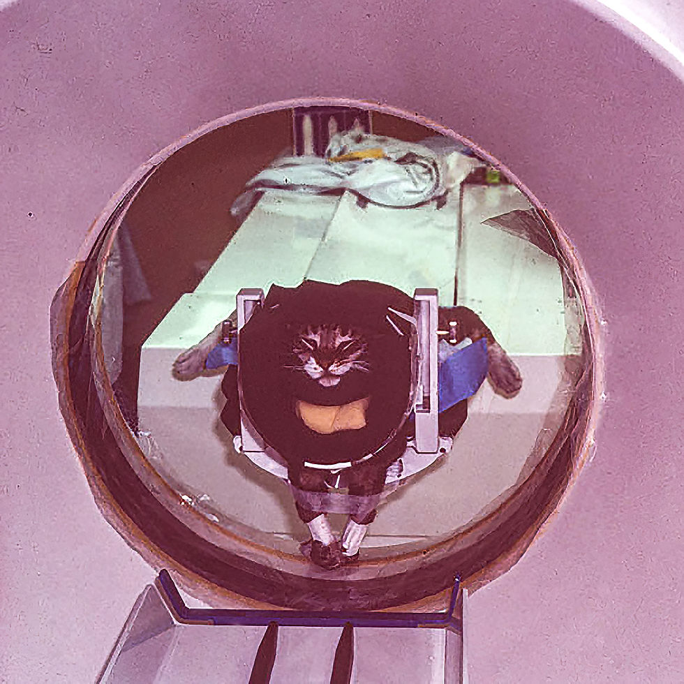
The earliest recorded CAT scan of a cat with a brain tumor was done in the early 1980s at the University of California San Francisco Medical (UCSF) Center. In a “cloak and dagger” exercise, Drs. John Fike, Chris Cann and I used the research scanner at UCSF to complete the first CAT scans of dogs and cats with brain disorders.
CT was a Game Changer for Feline Medicine
Before CT scanning, veterinarians relied on neurological examination findings, radiography, and exploratory surgery to diagnose brain tumors in cats. Cerebral angiography, being used in dogs, was not an option in cats due to the unique cerebral blood supply to the feline brain.
The advent of CT scans provided several advantages:
Detailed Cross-Sectional Imaging – Unlike traditional X-rays, which compress three-dimensional structures into a two-dimensional image, CT scanning allowed veterinarians to see precise slices of internal anatomy.
Non-Invasive Diagnosis – This reduced the need for exploratory surgeries, which were both risky and costly.
Better Neurological Assessments – Cats suffering from seizures, balance issues, or unexplained paralysis could be examined more thoroughly, helping veterinarians identify tumors, infections, or congenital abnormalities.
The First Feline Patients
Early veterinary CAT scans were primarily performed on cats suspected of having brain tumors. Since anesthesia was required to keep the animal motionless during the scan, early cases were often “high-stakes,” where the benefits of the procedure outweighed the risks. These scans confirmed conditions that previously had been challenging to diagnose, allowing for more targeted treatments.

Modern-Day Feline CT Scanning
Today, CT scans are a routine diagnostic tool in veterinary medicine, available not just at university hospitals but also in private specialty clinics. With advancements in imaging technology, modern veterinary CT scanners are faster, produce higher-resolution images, and allow for 3D reconstructions of a cat’s internal anatomy. Some newer machines, such as cone-beam CT scanners, provide even quicker imaging with lower radiation doses, making scans safer and more accessible.
From Curiosity to Common Practice
The idea of performing a CAT scan on a cat may have started as an amusing coincidence in name, but it has become a vital part of veterinary diagnostics.
What began as an experimental adaptation of human medical imaging has now saved countless feline lives.
Wouldn’t it be fascinating to see the expression on Sir Godfrey Hounsfield’s face if he knew that his groundbreaking invention had enabled veterinarians to scan actual cats?
References
1981 Fike JR, RA LeCouteur, CE Cann, CM Pflugfelder: Computerized tomography of brain tumors of the rostral and middle fossas in the dog. American Journal of Veterinary Research 42:275-281.
1981 Fike JR, RA LeCouteur, CE Cann: Anatomy of the canine brain using high resolution computed tomography. Veterinary Radiology 22:236-243.
1981 LeCouteur RA, JR Fike, CE Cann, VG Pedroia : Computed tomography of brain tumors in the caudal fossa of the dog. Veterinary Radiology 22:244-251.
1982 LeCouteur RA, JR Fike, RH Scagliotti, CE Cann: Computed tomography of orbital tumors in the dog. Journal of the American Veterinary Medical Association 180:910-913.
1983 LeCouteur RA, JR Fike, CE Cann, JM Turrel, JE Thompson, JF Biggart: X-ray computed tomography of brain tumors in cats. Journal of the American Veterinary Medical Association 183:301-305.
1984 Fike JR, RA LeCouteur, CE Cann: Anatomy of the canine orbital region-multiplanar imaging using CT. Veterinary Radiology 25:32-36.
1984. Fike JR, CE Cann, JM Turrel, RA LeCouteur, CM Pflugfelder: Relative uptake of low-and high-osmolality contrast media in CT of brain tumors. American Journal of Neuroradiology 5:413-417.
1985. LeCouteur RA, CE Cann, JR Fike: Computed Tomography. Chapter 44. In, General Small Animal Surgery, eds. IM Gourley & PB Vasseur, JB Lippincott Company, Philadelphia, p.989.
1986 Turrel JM, JR Fike, RA LeCouteur, RJ Higgins: Computed tomographic characteristics of primary brain tumors in 50 dogs. Journal of the American Veterinary Medical Association 188:851-856.


Comments